Blog 7/9

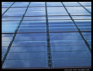
Today’s lecture greatly changed my interpretation of the collaboration between science and art. Though art has always been in collaboration with various forms of technology, the lecture opened my eyes to the complementary relationship that science and art share. By completing projects such as stained glass, artists and scientists present their collaborations to the rest of the public: scientists develop glass, searching for new methods of improvement and progression, and artists create captivating images and colors to transform the clear glass into a masterpiece. By completing their separate, yet equally important parts, scientists and artists produce beautiful pieces of stained glass that adorn many well-known locations.
The Weather Project
During our lab visits, we were taught about the new forms of energy sources being worked on. I was most interested about the possibilities of using algae as a means of energy. Algae would be extremely efficient because given the right conditions, it can double its volume overnight. Red tide is a phenomenon during which algae becomes so numerous that they discolor coastal waters. It can be caused by warm ocean surface temperatures, low salinity, high nutrient content, calm seas, and rain followed by sunny days during summer.
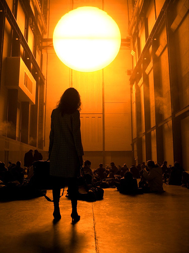
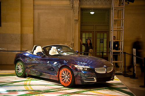
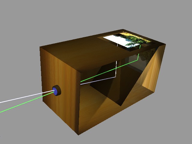
Another part of today’s activities that caught my attention was The Weather Project, created by Olafur Eliasson. In this exhibition, Eliasson mixed sugar and water to produce a mist, which he dispersed through a humidifier at the scene of the exhibition, London’s Tate Modern. While researching Eliasson, I noticed that he was commissioned by BMW in 2007 to develop the sixteenth art car for the BMW Art Car Project, showing another relationship between art and the science of engineering.
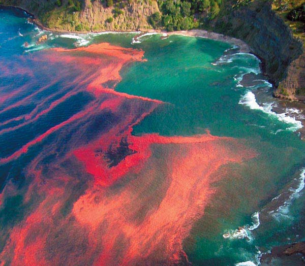
One of the BMW's from the BMW Art Car Project

Red Tide

Red Tide

Posted in Student Blogs Group X, Uncategorized | Leave a comment

July 9th Blog

Science, technology and art have always been something I have been intrigued to further advance my knowledge in. Listening to Giacomo Chiari incorporate all three realms was something remarkable. I was perplexed to realize how hand and hand all three subjects can be alike. Learning about the technology available to restore art using scientific data was extremely fascinating. This lecture was surely not cut and dry, and fused the whole audience with interest.

As if my mind wasn’t ecstatic already, I was flabbergasted to be the one chosen to put my hair under the experimental electron microscope. This piece of machinery was very user friendly and allowed even I to control it with ease. Of course, you do give up focus for easy access and petiteness. My hair was immensely interesting to look at under such a device. It showed detail that I have never seen before, and if that wasn’t enough I even got to measure the diameter of my hair (approx. 75 microns). Today was truly something I will remember for the rest of my life, and using a touchscreen electron microscope, is no ordinary luxury, and I feel extremely fortunate to be one of the select few who get to enjoy that experience. I hope more days are soon to come with this excitement. All in all, today was packed with fascinating labs, lectures, and activities that kept me not just occupied, but satisfied. Watching so many college students and professors, has inspired me even more to carry out research immediately following this program.

http://www.getty.edu/conservation/science/current.html

http://www.labx.com/v2/adsearch/detail3.cfm?adnumb=389990

http://www.bruker-axs.de/introduction3.html

http://newsroom.ucla.edu/portal/ucla/ucla-researchers-create-polymer-72064.aspx

http://www.cnsi.ucla.edu/arr/paper?paper_id=196881

Posted in Studnt Blog Group Y | Tagged | Leave a comment

7/7

Today, we were introduced to some of the similarities between art and science as we listened to our morning lecture on Visualization. It began with some ideas about how both art and science can serve to be useful tools for each other. The question “How do artists and scientists produce images?” was asked. Perspective was a common theme in both the arts and sciences. Rays were discussed as being an example of a scientific manifestation of one-point perspective. The artist Jon Von Ike was also mentioned as an artist who used optics in his paintings. I have always found this painting interesting and complex and now I know why. Multiple, hidden ideas permeate the painting and allow for a much more complex and interesting outcome. Not only paint was used but also the idea of the bending of light in a mirror was in mind when Jon Von Ike painted this piece. The idea of perspective was continued when the idea of “nano” was introduced. This scaling down of an image allows one to look at an image with a whole new perspective. This interpretation of the minute details of something can also be seen when analyzing pieces of art, poetry, and music. There can be found a deeper meaning in most forms of art when looking at it from different perspectives. The content which is able to be seen on the surface is not always the most important piece of information to be taken from the art as is with the nano particles seen in this developing science of nano technology. I found this first lecture to be stimulating and thought provoking and I am excited to see what other connections are going to be made between art and science later this week.

http://en.wikipedia.org/wiki/Jan_van_Eyck

http://brightbytes.com/cosite/what.html

http://www.unl.edu/CMRAcfem/em.htm

Posted in Student Blogs Group X | Tagged | Leave a comment

JULY 8, 2009

Today a very cute old man came in to talk about NanoMedicine. Am I allowed to say he’s old? Is that offensive? Well I mean it in a superly awesome and good way. Anyways, he discussed the use of engineered molecular systems and molecular knowledge of human body to maintain. The different tools made with nanotechnology could be used for diagnosing diseases, biological materials, drugs and drug delievery. He also talked about nanofibers which could be used as biosensors. The nanofibers are made of polyanaline fibers, which will come into contact with vapors which then conducts electricity.060310_nano_fibers_021

Look at those skinny little nanofibers!

vault_jmol
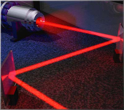
The power of the current varies according to the pH of the vapors. The most interesting thing that he talked about were the vaults. The vaults are made of three proteins and a small RNA. There is the Major Vault Protein (MVP), the TEP1 and the VPARP. Unfor.tunately, the true function of these unusual vaults are unknown. Personally, I think that the vaults are probably useless like the vestigial organs in the human body (i.e. tailbone and appendex). Even though the scientists say they are going to use them as drug capsules to treat cancer, I feel like they will not be effective enough.  They might be helpful in addition to chemotherapy, but they are probably not strong enough by themselves.

My favorite thing today was the zero wave function. I loved the elegant and smooth flowing movement of buckminster balls. I liked how I was able to make them larger and smaller with my hands.

My least favorite thing was having someone constantly taking pictures and filming me. It made me feel uncomfortable and self concious about my fat and ugly grossness. But thats ok I guess, because that’s her job and all.

weber13b

Posted in Student Blog Group Z | Leave a comment

Day #3: Art, science and nature

Nanotechnology is one of today’s fastest growing fields. It has enormous potential. Nanotechnology can either revolutionize our world in ways that no one can possibly predict or it can lead to our destruction if it is not properly controlled and regulated. Nanotechnology is powerful because of it’s small size, but that size, thousands of times thinner than a human hair allows nanomaterials to easily pass into human cells, where they can possibly disrupt cell functions or cause the destruction of those cells. Today nanotechnology has progressed so far that we can encounter it and interact with it almost everyday, in many cases without our knowledge.  The United States currently has a “innocent until proven guilty” policy dealing with many new materials and technology. The dangers of using nanotechnology are not fully known and will not be for many years to come. Until then, the use of nanotechnology and the research of it will be increasing at an ever increasing rate. If today’s trends continue into the future society could be entirely immersed in nanotechnology by the time that it’s dangers are fully realized.

However, Berkeley, California has taken the initiative and has placed nanoparticles on it’s hazardous materials list. It’s move was based on years of re search into nanotoxicology, a topic covered today by Hilary Goodwin.

ucla-sci-art-018

nd-silver-particles1

Today, nanosilver particals are in wide use in athletic clothing because of silver’s antibacterial properties. One of the dangers it poses is that those nanosilver particles have been shown to fall out of the clothing and into the water. Silver’s antibacterial properties affect not only harmful bacteria but beneficial bacterial as well. As nanosilver particles leak into the enviornment they will adversely affect it. So far, the effects have been inconsequential, but with continuing pressure, even the Earth’s mighty envoirnments, like the stain resistant lab coats demonstrated to us, will eventually yield.

Hydrophobic and stain resistant lab coat demonstration

Hydrophobic and stain resistant lab coat demonstration

http://thetyee.ca/News/2008/04/07/NanoParticles/

http://www.sfgate.com/cgi-bin/article.cgi?file=/c/a/2006/11/24/MNGP9MJ4KI1.DTL

http://scitation.aip.org/getabs/servlet/GetabsServlet?prog=normal&id=BJIOBN00000200000400MR17000001&idtype=cvips&gifs=Yes

http://www.dmoz.org/Science/Technology/Nanotechnology//

http://www.vega.org.uk/video/programme/3

Posted in Student Blog Group Z, Uncategorized | Tagged | Leave a comment

Vaults still remain a mystery

07.08.09 UCLA-Until the mid 1980’s the Vault wouldn’t be truly noted until a study conducted by Professor Leonard Rome who, at the time, was studying the birth and death of proteins. Even today this protein made structure, comprised of three major proteins and a small strand of RNA, remains to hold no known function. Today we were fortunate enough to have Professor Leonard Rome, himself, come in and speak on the subject. As one would expect, Leonard Rome, Dean of UCLA’s Medical, was truly just a brilliant man. Yet, perhaps more valuable then brilliants itself was his passion. Professor Leonard Rome knows his subject inside and out, literally.

Named Vaults due to their structure, these tiny capsules found commonly in all red blood cells of the majority of eukaryotic cells, consist mostly of MVPs (Major Vault Proteins). This makes up around 70% of he Vaults themselves. Some of the other major parts of the Vault cell particles is Cap Proteins and vRNA. And despite the developed information towards the structure of the vaults still no information at this time is known regarding their function.

I personally cannot come up with an explanation for what or how these vaults may serve as. A true guess and wild speculation would be that these cell particles have the purpose of resistance. The coding of the vRNA is very simplistic compared to the giant coding of normal RNA which suggests a much more simplistic function. Also it could be guessed that there could probably be an extra copy hidden in the nucleus of the cells as a blueprint for building the cells. However, if in the case that the nucleus is destroyed or damaged the cell may be able to use the vault as a backup resistance in hopes of not a complete shutdown.

One other note I’d like to add to this blog how inspiring it was to see the true passion Professor Leonard Rome had for his Vaults. He stated his passion for his subject popped up in his head as often as food, water, and sleep pops into the average persons head. This to me, serves as a true inspiration because without a true passion like Professor Leonard Romes would never solve something as mysterious as the Vault cell most likely because the outlook of finding out its actual function looks very bleak. I hope Professor Leonard Rome keeps on with his wonderful studies.

http://vaults.arc.ucla.edu/static/bkidspg/bk-rna

http://en.wikipedia.org/wiki/Vault_RNA

http://users.rcn.com/jkimball.ma.ultranet/BiologyPages/P/Proteins.html

http://www.usoe.k12.ut.us/CURR/science/sciber00/7th/cells/sciber/orgtable.htm

http://www.vaults.arc.ucla.edu/static/scipg/sci-history

Posted in Student Blogs Group X | Leave a comment

Wednesday the 8th

Today we talked about how nanotechnology can help with cancer. Right now we use chemotherapy, which is a form of radiation, to fight the cancer cells. The problem is that it kill ALL the cells. Doctors basically try to kill the cancer before they kill the whole patient. Obviously this is problematic so now they are designing new ways to target just the cancer cells. One way to do this is to target cells with low PH because cancers cells have been found to have a lower PH than the rest of the body. Another way they do this is by having medicine be spread throughout the body in nanoparticles that release the medicine when light reaches them. This was scientists can light up just a tumor and release the drugs right there.

In addition to looking deeper into cancer cells, we went down into a lab with a group of scientists who are turning phones and webcams into devices that could save lives. They modify them to take pictures of a blood sample easily and quickly. This way people who do not have doctors nearby, like those in developing countries, can get diagnosed based on the red blood cells in their body by sending the picture to a doctor. The doctor can even text the diagnosis back to the patient.

Red Blood Cells

I found this research very amazing because they already have working examples of the entire system. It it innovative and solves a real problem that can help people today.

http://www.jumpintotomorrow.com/template/index.php?tech=245

http://www.youtube.com/watch?v=AzX0Vx4Aibk

http://dailybruin.com/stories/2007/may/11/ucla-research-fuels-cancer-therapy/

http://www.youtube.com/watch?v=-wFYxx8C2os

http://www.youtube.com/watch?v=4wXjIxWo94c

Posted in Student Blogs Group X | Leave a comment

Tuesday the 7th

In the morning we saw a video called “Imagining the 10 Dimensions” which I found interesting. It was not based fully on the accepted version of string theory but it was a cool way to think regardless. Here is Part 1 and Part 2 of the video.

After the morning intro video we looked at the microscopes in the basement floor of the building. We saw million dollar electron microscopes as well as more traditional light microscopes. One of them (a confocal microscope, I think) took pictures in slices and then built them into a digital 3D image; it is the only one that can do that in the entire US. Using the electron microscope we zoomed in a million times to see tiny particles called Vaults. These were discovered right here at UCLA and have no known purpose yet. The guy showing them to us used liquid nitrogen to freeze the vaults and even splashed it at us. He also had a cool accent.

The scanning electron microscopes looked like something out of the movies. They built every part and assembled them in the lab because they are not even sold anywhere in the world. The actual microscope part is very small but the housing is large so the scientists can move samples around in a vacuum.

Posted in Student Blogs Group X | Leave a comment

Blog 2 July 8

self-cleaning-windows

There was a time when nanotechnology was a term that nobody knew. Some people even today think the iPod nano is nanotechnology. However there is an observable pattern that nanotechnology is becoming more and more part of pop culture.

1984 
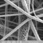
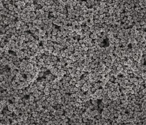
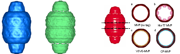
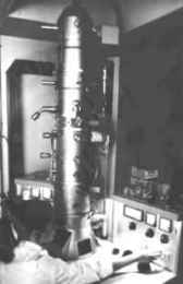
Now days, nanotechnology is being used in everyday objects that make people’s lives convenient such as self-cleaning windows and odorless socks. The significant truth is that these achievements were thought to be impossible in the past, but now are part of reality. By assuming this, scientists predict that contraptions that appear unachievable today may become feasible in the future. Some of these inventions have been part of movies which have later become reality such as “1984.”

In the modern movie “I, Robot”, a detective named Del Spooner lives in Chicago in 2035 with great contempt for robots, which in this world are universal. Detective Spooner’s immense disdain began when he was involved in a car crash. A truck driver fell asleep during his drive and smashed Spooner’s car into another, causing both to fall into the ocean. While sinking, a robot, which was nearby and saw the accident, dove and swam its way toward the demolished vehicles. Although it was saving Spooner, he yelled to save the girl in the other car. However the robot calculated Spooner had the highest probability of survival; thus the young child unfortunately drowned.i-robot-arm

Due to this mishap, Detective Spooner lost his left arm and lung. As a result, a doctor replaced them with a robotic prosthetic arm and lung, which later in the movie becomes very useful. Although surgeries like these are ridiculously difficult, it may become a simple task once nanotechnology procedures are completed and polished.

http://en.wikipedia.org/wiki/I,_Robot_(film)

http://chucksconnection.com/irobot.html

http://www.moviemistakes.com/film4390/questions

http://www.webmd.com/lung-cancer/lung-surgery-thoracotomy-for-lung-cancer

http://www.zyvex.com/nano/

Posted in Uncategorized | Tagged | 1 Comment

Blog 1 July 7

light-microscope

Just when scientists thought they explored the whole world, numerous more arrived. During the Renaissance, the light microscope was developed which was the first instrument that enabled the human eye to observe enlarged images of infinitesimal objects. This incredible invention brought fascinating detail of the tiny worlds invisible to the naked eye. About 1950, Dutch spectacle makers, Zaccharias Janssen and his son Hans, were experimenting in the Netherlands with several lenses in a tube. They discovered that the nearby objects appeared greatly enlarged.

Years later Anton van Leeuwenhoek, of Holland the father of microscopy, taught himself methods for grinding and electron-microscope
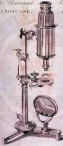
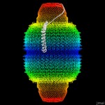
polishing tiny lenses with great curvature as an apprentice at a dry goods store where magnifying glasses helped count threads in cloth. These eventually led to the building of his light microscopes and famous biological discoveries which made him well known. This man was the first to observe and describe bacteria, yeast plants, drops of water, and blood circulation. Anton’s discoveries and studies ended up being reported as findings in over a hundred letters to the Royal Society of England and the French Academy.

In the 1930’s, the first electron microscope was introduced by co-inventor Germans Max Knoll and Ernst Ruska. They were awarded half of the Nobel Prize for Physics in 1986 for this invention. In this new kind of microscope, electrons are speeded up in a vacuum until their wavelength is extremely short. Beams of these amazingly fast electrons are focused on a cell sample and are scattered or absorbed by the cell’s organs. This forms an image on electron-sensitive photographic plate of its structure.

cellmicroscope-sample-mayb

http://www.microscopesguru.com/when-was-the-microscope-developed/

http://inventors.about.com/od/mstartinventions/a/microscope.htm

http://www.brianjford.com/whistmic.htm

http://www.microscope-microscope.org/basic/microscope-history.htm

http://campus.udayton.edu/~hume/Microscope/microscope.htm

Posted in Uncategorized | Tagged | Leave a comment