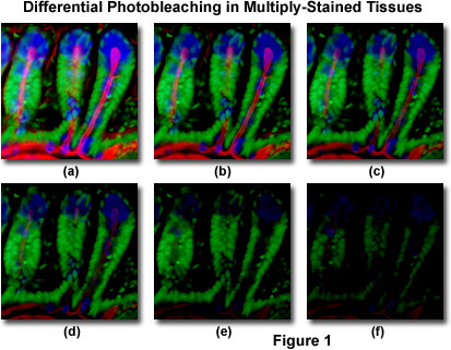
The visits and exploration of the core labs this afternoon helped me to really see how the collaboration of scientists and artists can result in amazing works of art and developments in science. I was especially interested in the use of fluorescent and quantum dots to light up or label parts of a sample. The fluorescent labeling is more general and may react with many different cellular components within a sample. The use of quantum dots in labeling is quite different from the fluorescent technique. A quantum dot is a semiconductor that is used in labeling samples. It is a good technique because it is resistant to photobleaching. Photobleaching is the destruction of a flourophore or part of the molecule. This can cause complications when viewing the labeled part of the sample. These properties make it a useful technique when tracking hematologic cells. The department of medicine in UCLA is using this technique to help track human leukemic, bone marrow, and cord blood cells. By using quantum dots for labeling the hematologic cells the cells can be track up to four cell divisions. I believe this would help determine how fast the cancer is spreading and help in treatment. Hopefully it will also lead to new types of medicines that can hinder the spreading of the cancer. A really amazing fact is that the die can kept a cell labeled for up to two weeks. This allows scientists to really follow the cells through the cell cycle and study how it affects the overall speed at which the leukemia spreads.


http://en.wikipedia.org/wiki/Fluorophore
http://jco.ascopubs.org/cgi/content/abstract/23/10/2264
http://en.wikipedia.org/wiki/Photobleaching
http://en.wikipedia.org/wiki/Quantum_dot
http://www.ncbi.nlm.nih.gov/pubmed/17027955
