Today we learned about different methods in nanoscience research. The day opened with a presentation by Rita and Gau that focused on visualization, art, and science. Visualization is the technique of creating an image, diagram, or other visual to express an idea. I knew there were multiple methods of producing an image in art such as oil paint, sandstone, and marble, but I never really thought about the different methods in science. The presentation and lab visits today exposed me to this new idea.
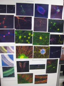
Images produced using microscopes
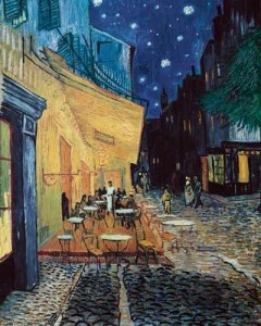
Image produced using paint
During the powerpoint, Rita and Gau stressed that in visualization, art inspires science, then science inspires art, and so on. For example, Charles Darwin, a scientist, was influenced by art, and he later influenced artists. I found it interesting that the connection between art and science dates back to the 1800s with Darwin and even further back to the Italian Renaissance. The presenters also emphasized that there are various ways of interpreting something, and the way we see something molds our views on it. This concept caused me to open my mind and be more aware of the way I look at things.
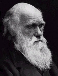
Darwin was inspired by art, and he inspired artisits
The three lab visits today focused on different methods of visualization in science. I enjoyed learning about different types of microscopes and how they work. For example, our first lab was “Electron Microscopy.” An electron microscope uses a beam of electrons to light up the sample and create a magnified image. After learning about the process of preparing a specimen for this type of microscope, we were able to play with the electron microscope and adjust the magnification, light, position, etc. This hands-on experience helped me to grasp the concepts of the day.
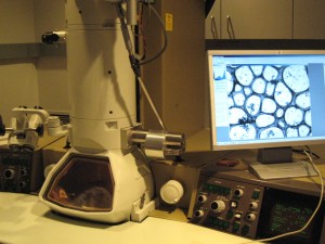
Using electron microscope to look at sample from liver cells
http://royalsociety.org/page.asp?id=1213
http://www.nsf.gov/news/overviews/nano/index.jsp
http://vlib.iue.it/carrie/texts/carrie_books/gilbert/07.html