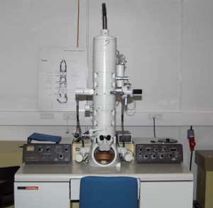
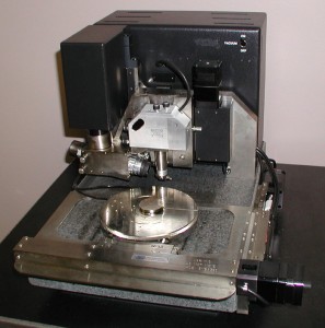
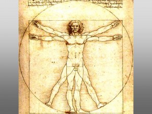
The intertwining of art and science is an interesting thing. Many of the early artists incorporated useful methods of producing a 2-D image from a 3-D model, such as using manageable squares and a ray of light (1-D perspective). One of the first methods of translating a 3-D to 2-D was the use of a camera obscura and what is truly amazing is that many of our cameras today incorporate this same basic function only today’s, of course, are more advanced. Louis Pasteur’s and Joseph Lister’s works are the foundation of the modern medicine and applied every single day. The smooth enamel on teeth, superficially smooth, are actually rods, called crystals of appetite, that lean against each other in the same direction. There are many types of microscopes incorporated in today’s research and analyzation of experiment results. The scanning electron microscope, SEM, uses an electron beam focused into a very fine probe at the surface of a solid specimen. A transmission electron microscope, TEM, also uses an electron beam, but the electrons go through the sample and is received by a detector on the opposite side, thus leading to a higher resolution than SEM. The atomic force microscope utilizes a cantilever with the tip at the end scanning over the surface. This allows for manipulation of the atoms. In the future, there will be further advancements in microscopy that allow for subatomic viewing and manipulation of subatomic particles. Many ideas emerged during the Italian Renaissance, including the idea of humunculus in which humans lived in the head of the sperm before birth and the egg played no part in reproduction. One of the most renowned artistic scientists was Leonardo Da Vinci. His brilliant works included artistic aspects and also scientific aspects of life. He magnificently created and worked on hydrodynamics, aerodynamics, nature, anatomy, inventions, and paintings. The supposed immense gap between art and science is rapidly closing and they are becoming closer and closer to being one.


