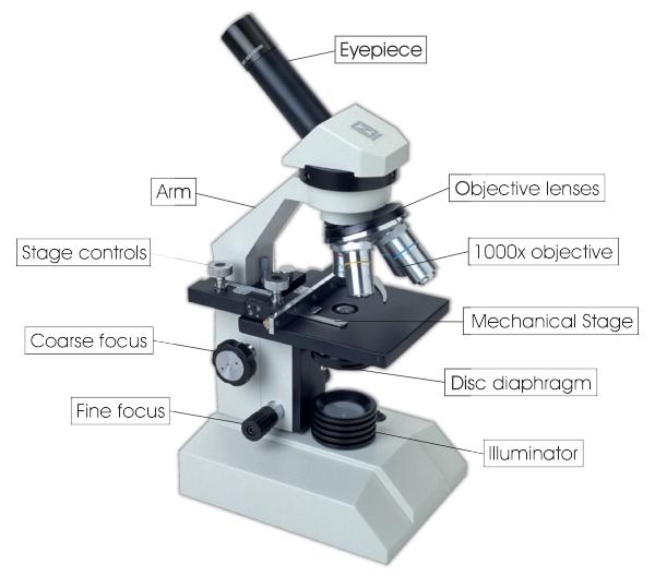
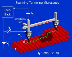
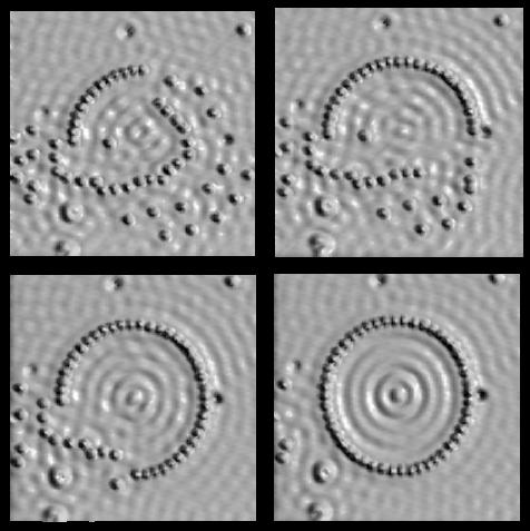
Today I began my understanding of the artscience program. The motif for the day was magnification and microscopy. We saw multiple microscopy labs and met lots of people who worked with imaging and magnifying devices all day. Most of what I saw I had heard about before. We saw a normal light microscope and even an electron microscope, however we were also shown a machine I had never known existed. The machine in question is called a scanning tunneling microscope which uses a very sharp needle, (sharpened down the molecular level.) to actually interact with the surface one is intending to visualize. Using intermolecular and even atomic forces the STC can map the “topography” of a surface at the molecular scale. This machine also demonstrated to me the entry of art into microscopy. The STC is different from other microscopes in that it can be used to interact with the surface of the material. The sharp point of the STC can be used to pick up and move atoms into shapes and patterns by jolting atoms with electrical shocks and causing them to stick to the carbon nanotube that is the microscopes tip. This technique can be used to rearrange atoms into a patern. This manipulation of atomic structure can be captured by a camera. The pictures produced are a new form of atomic art that owes its existence to advances in microscopy. I began to appreciate, through this new type of art, what it means to link art and science. Today’s activities helped me further understand what this program is about.
links:
http://dpmc.unige.ch/gr_fischer/localprobe.html
http://en.wikipedia.org/wiki/Scanning_electron_microscope
http://medicalphysicsweb.org/cws/article/newsfeed/39549
http://micro.magnet.fsu.edu/micro/gallery.html
http://www.mos.org/sln/SEM/