Day 1: Top-Bottom; Bottom-Up
Macro. Micro. Large. Small. This is what differentiates the visual world from the invisible.

Today, we learned about the different microscopes used by scientists to accommodate the growing field of nanotechnology. Among these include compound microscopes, electron microscopes, field ion microscopes, and scanning tunneling microscopes. Truly, the human eye has evolved past its biological confines to venture into the nanosphere. No longer do we have to depend on the limitations of nature’s given spectrum.
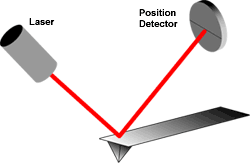
While the light microscope has been a fixture in the scientific society, the visible light spectrum has proved too constricted for nanotechnology use. Therefore, we were introduced to the atomic force microscope, a cutting-edge technology that relied on the pressure exerted by the atom on the end of the tip of the probe (cantilever) to scan the surface of the sample in order to dictate its size and shape. Like a paintbrush, the pressure applied and the contact between probe and “artwork” determines the outcome of the vision.

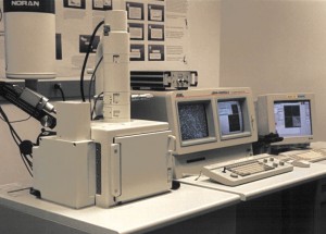
Serge then showed us a variety of machines that CNSI had in the basement that included a centrifuge and a scanning electron microscope. The scanning electron microscope had the ability to magnify the sample up to a few hundred nanometers. I find it ironic that what is closest to us, ie. the nanospectrum, is the most foreign and jarring to visualize. Perhaps that is the charm of art as well. That we are enabled to see what is in front of us but cannot without the instrument of science.

Links
http://www.cnsi.ucla.edu/
http://www.britannica.com/EBchecked/topic/93144/cantilever
http://www.mos.org/sln/SEM/
http://www.nanoscience.com/education/AFM.html