This day was an extremely eye opening one.
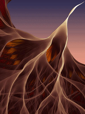
The day started with our daily lecture on the thin lines between art and science and helped dissolved the barriers even more. We learned about a variety of art that had truly broken the line between art and science. 
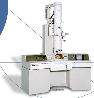
We separated into our groups and went off to do our lab activity for the day. Today was different types of microscopes and there was a lot of information that needed to be covered. The first lab we visited showed us an extremely high powered light microscope. After a short lecture on how the microscope worked the professor explained to us that this microscope was an extremely useful too because this microscope could look at live tissue rather than an electron microscope which can only view dead things. Electron microscopes may be inconvenient because they can only view dead material, but they make up for this disadvantage by having extreme magnification powers.

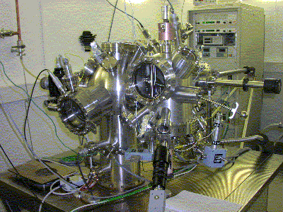
After viewing the two other incredible microscopes we moved on to the last and most surprising of the microscopes, the electron tunneling microscope. This microscope has an extremely tiny needle that is attached to a structure with runs it along a moving plate where the sample is held. This needle head “feels” the surface of the atoms and gives a topographic readout of the atomic structure of the sample material. This microscope can also be used to manipulate atoms into forms scientists want.

Today was and insightful and extremely fun day.
http://en.wikipedia.org/wiki/Scanning_tunneling_microscope
http://en.wikipedia.org/wiki/Electron_microscope
http://en.wikipedia.org/wiki/Optical_microscope
http://en.wikipedia.org/wiki/Microscopy
http://www.ruf.rice.edu/~bioslabs/methods/microscopy/microscopy.html


