Ah! Okay so where are we currently. First day and we got introduced to a trillion long words that I can hardly remember. In all science research laboratories contain microscopes! Woo microscopes. We were introduced to microscopes. They all had their different function and each one got better than the previous. Let’s see we had, scanning tunneling microscope, Electron microscope, transmission electron microscope, atomic force microscope. Woo. Joyful. So what is the difference of all these microscopes.
Let’s begin with the first one. The scanning tunneling microscope.  This type of microscrope shows 3-D images from the sample. Cool right? If you want to know how deep your specimen is, or how sticky or any other thing that comes to mind. This would be the right microscope for you to use. The tip of the microscope is one atom. Cool right? STM works mostly with conducting materials but it can also work with the organic ones.
Our next microscope is the electron microscope. Except there are a lot of different electron microscopes so what is the difference between all these? Well first, an electron microscope uses a beam of electrons!! in order to produce a high resolution image. Why an electron microscope? Well these microscopes can produce images that are 200 million times with the use of electromagnetic radiation. Cool right?
Different type of scanning electrons include: Transmission Electron Microscope, Scanning Electron Microscope, Scanning Transmission Electron Microscope. A lot of microscopes. Ah! Okay so what is the difference between all of these? Why cant we use one and make it amazing to view all the types of images possible. Hm… not sure. But

Lets see, the first microscope is the Transmission Electron Microscope or TEM. This microscope works like a slide projector where you put the image down and it gets shone at by a bunch of tiny little electrons. What we see is what is reflected by the light we see. The TEM works something along the lines of the image to the left.
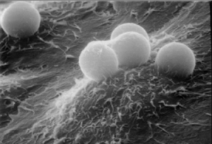
Next we have the Scanning Electron Microscope. This microscope also uses electrons instead of light to produce an image. The SEM has a large depth field so you can see more of your specimen at once. the SEM uses electromagnets instead of regular lenses which gives the researches more options when it comes to zooming in to their specimen.
We also have a Scanning Transmission Electron Microscope. The electron optics focus on one spot and allow for it to scan a raster. With this microscope it is possible to obtain atomic resolution images. Amazing right?
Links:
http://nobelprize.org/educational_games/physics/microscopes/scanning/index.html
http://en.wikipedia.org/wiki/Electron_microscope
http://www.unl.edu/CMRAcfem/temoptic.htm
http://www.purdue.edu/REM/rs/sem.htm
http://en.wikipedia.org/wiki/Scanning_transmission_electron_microscopy