By Lily | Published:
July 8, 2009
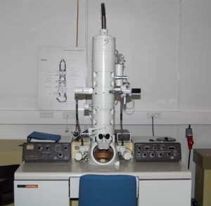
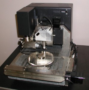
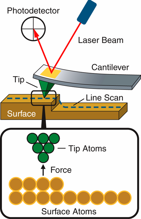
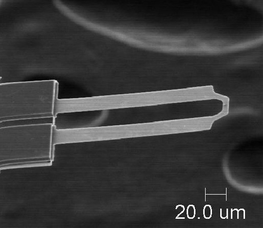
Today we went to the microscopes labs and learned bout the different types and got to look through them. A few types included compound, electron, field ion (visualize atom) scanning tunneling function (structure). Three types: the Scanning Electron Microscope (SEM), the Transmission Electron Microscope where the electrons go through the sample and detectors receive electrons on the other side; which has higher resolution than SEM, and the Atomic Force Microscope where a cantilever with a tip at the end is scanned over the surface. The tips balanced so that the gravity force is attracted/ makes it go down, and Van Der Waals force which detracts it/makes it go up.
We learned about top- bottom (macro) like when a sculptor takes a piece of marble and chizzes it down to get what he wants; for example pyramids and arches, self assembly. Lithography (printing) and bombardment (sculpting) Bottom-up (micro) is building atom by atom.
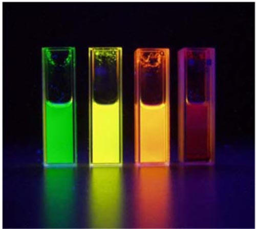
We also learned about quantum dots (qdots) which are materials that’s size is dependant to the electronic confinement.
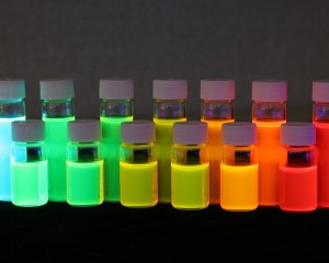
The color change is accordance to size, where the electron moves faster because there’s more energy and less space. We also talked about teeth enamel, which has the hardest surface in the human body. It’s made once and can’t be remade so once there’s holes and cavities forming you can’t bring back that enamel. Artists produce an image, record and communicate their ideas. Examples: oil pan, marble, sandstone, celluloid, and cells. Microscopes get an image and spectroscopies gets data and infer an image. The light microscope was created in 1609 and the x-ray was in 1895. Learned that x-ray diffraction diffracts at 2x incident angle. We got to go into the labs and look through some types. A cool one was the light microscope where you could pic a specific color and only show that part of the cell and the other parts were not visible.
http://www.southwestschools.org/jsfaculty/Microscopes/types.html
nano.tm.agilent.com/blog/page/3/
https://www.llnl.gov/str/Lee.html
http://www.dentalfind.com/glossary/tooth-enamel.html
http://www.chemguide.co.uk/atoms/bonding/vdw.html
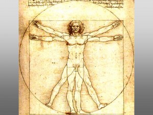
The intertwining of art and science is an interesting thing. Many of the early artists incorporated useful methods of producing a 2-D image from a 3-D model, such as using manageable squares and a ray of light (1-D perspective). One of the first methods of translating a 3-D to 2-D was the use of a camera obscura and what is truly amazing is that many of our cameras today incorporate this same basic function only today’s, of course, are more advanced. Louis Pasteur’s and Joseph Lister’s works are the foundation of the modern medicine and applied every single day. The smooth enamel on teeth, superficially smooth, are actually rods, called crystals of appetite, that lean against each other in the same direction. There are many types of microscopes incorporated in today’s research and analyzation of experiment results. The scanning electron microscope, SEM, uses an electron beam focused into a very fine probe at the surface of a solid specimen. A transmission electron microscope, TEM, also uses an electron beam, but the electrons go through the sample and is received by a detector on the opposite side, thus leading to a higher resolution than SEM. The atomic force microscope utilizes a cantilever with the tip at the end scanning over the surface. This allows for manipulation of the atoms. In the future, there will be further advancements in microscopy that allow for subatomic viewing and manipulation of subatomic particles. Many ideas emerged during the Italian Renaissance, including the idea of humunculus in which humans lived in the head of the sperm before birth and the egg played no part in reproduction. One of the most renowned artistic scientists was Leonardo Da Vinci. His brilliant works included artistic aspects and also scientific aspects of life. He magnificently created and worked on hydrodynamics, aerodynamics, nature, anatomy, inventions, and paintings. The supposed immense gap between art and science is rapidly closing and they are becoming closer and closer to being one.
Links
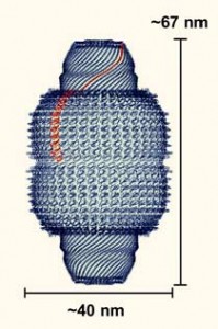
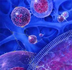
Nanotechnology consists of structures and technology relative to a nanometer. A nanometer is a unit of measure equal to one billionth of a meter. Many forms of technology exist today that utilize nanotechnology already, i.e. nano-silver lined socks, nano-coated “self-cleaning” cement, nano-zinc in suntan lotion, and many more in cosmetics. Above all, Biomedical applications of nano technology succeed any others in importance, and usefulness in our society. Dr. Rome is a lead researcher in the studies of Cancer treatment by using nanotechnology to distribute medicine throughout the body, targeting certain cells. Rome’s team bases their research on Vaults, capsules in the body that can travel through the blood stream to target cancer cells. These capsules are naturally found throughout the human body. Researchers have determined no definite use for the vaults in the human body, and have speculated that it is safe to utilize them for cancer tumor treatment. The Vault is composed of 3 proteins and a small RNA. The proteins are MVP (Major Vault Protein) and VPARP, TREP1. The small RNA is called Vault RNA. This vault is only 41 by 72 nanometers, and therefore it is easy to attach to cells. The vaults used for medicinal delivery are composed of adding MVP to DNA and inserting it into an insect cell. After that, the Vault assembles itself and is ready for use. These vaults have only been tested in Petri dishes, and will soon be ready for animal testing. In theory, if everything goes accordingly, then these vaults could be ready for use on humans in about 2 years. This would bring out a new era of medicine, and medicinal treatment. The theory could not only be applied to cancer, but almost every disease plaguing humans today. Nanotechnology could reinvent the future medicinal world as we know it.
http://www.foresight.org/Nanomedicine/
http://en.wikipedia.org/wiki/Nanomedicine
http://www.physorg.com/news3260.html
http://www.scientificamerican.com/article.cfm?id=nanomedicine-targets-cancer
http://www.nanoscienceworks.org/articles/nanomedicine-project-targets-prostate-cancer/?searchterm=Nanomedicine%20Cancer
Posted in Uncategorized | Tagged Y |
Today I was inspired by the topic of nanotechnology in medicine and all the different research being done to find cures for horrible diseases that were once deemed as incurable such as cancer, Parkinson’s, or sickle cell. The idea of nanobots, or robots on a nano scale, is a great example of how the sciences and arts are coming together and becoming less distinct. I believe artists or engineers are needed in the designing and structure of these machines while the sciences develop the theory and how it will work. The future of nanobots is that they will have the capacity to sense their environment and adapt to it. This leads to the question of once we have such advance technology what are some harmfully things it could lead to. Plus, is there going to be restrictions on what this technology can be used to do. Thinking about the affects of this technology reminded me of the series The Uglies, by Scott Westerfeld which has to do with using technology to take away the flaws of human life. Also in the story there is the technology of placing skin grafts on a wound to heal it, which is what some researchers are now using to heal wounds or holes inside the body or most commonly burns. There are two different types of skin graft. The more common one is where a thin layer of skin is removed from a healthy part of the body, donor section, and used for the skin graft. The second type is more of a risk; it is a full thickness skin graft, which involves cutting skin away from the donor section. This is more of a risk in terms of the body accepting the skin graft and not rejecting it from the body or killing it. The risks of this procedure are bleeding, infection, loss of the graft, or nerve damage.


http://en.wikipedia.org/wiki/Nanobot
http://www.nanobot.info/
http://www.amazon.com/Extras-Uglies-Scott-Westerfeld/dp/1416951172/ref=sr_1_5?ie=UTF8&s=books&qid=1247107522&sr=1-5
http://en.wikipedia.org/wiki/Nerve_damage
http://en.wikipedia.org/wiki/Skin_grafting
Posted in Uncategorized | Tagged group y |

 In the first two days of being introduced to nanotechnology, as we reviewed, amongst other things, the correlation between the development of microscopes, and their corresponding aid in scientific progress. A couple of the microscopes that we learned of and caught my interest were the atomic force microscope, and the scanning electronic microscope. The atomic force microscope is amazing in that it can reach down the fractions of nanometers. It functions through a cantilever with a tip on its end that scans the surface of an object. The tip in the cantilever is given a constant amount of pressure, and the image displayed on the microscope’s screen is determined on the amount of the distance the tip is displaced as it scans the surface. Second, the scanning electronic microscope works by the use of electrons. Electrons are shot at a sample, and they bounce off to a detector which draws a picture of the sample based on the intensity the electrons travel in. One of the scientific discoveries discussed in the lectures that surprised me the most was the radical color, and behavioral changes that any material undergoes as the corresponding material is viewed in an extremely small portion. These changes that occur are due to a process called quantum confinement, where electrons in a certain material are forced into a smaller area. This process additionally shortens the wavelengths of the given material which changes its color in the spectrum according to the size of the container the electrons are confined in.
http://www.ringsurf.com/online/2003-structures.html
http://www.mos.org/sln/SEM/
http://www.nanoscience.com/education/AFM.html
http://www.purdue.edu/REM/rs/sem.htm
http://www.physlink.com/News/101303QuantumWires.cfm
The visits and exploration of the core labs this afternoon helped me to really see how the collaboration of scientists and artists can result in amazing works of art and developments in science. I was especially interested in the use of fluorescent and quantum dots to light up or label parts of a sample. The fluorescent labeling is more general and may react with many different cellular components within a sample. The use of quantum dots in labeling is quite different from the fluorescent technique. A quantum dot is a semiconductor that is used in labeling samples. It is a good technique because it is resistant to photobleaching. Photobleaching is the destruction of a flourophore or part of the molecule. This can cause complications when viewing the labeled part of the sample. These properties make it a useful technique when tracking hematologic cells. The department of medicine in UCLA is using this technique to help track human leukemic, bone marrow, and cord blood cells. By using quantum dots for labeling the hematologic cells the cells can be track up to four cell divisions. I believe this would help determine how fast the cancer is spreading and help in treatment. Hopefully it will also lead to new types of medicines that can hinder the spreading of the cancer. A really amazing fact is that the die can kept a cell labeled for up to two weeks. This allows scientists to really follow the cells through the cell cycle and study how it affects the overall speed at which the leukemia spreads.


http://en.wikipedia.org/wiki/Fluorophore
http://jco.ascopubs.org/cgi/content/abstract/23/10/2264
http://en.wikipedia.org/wiki/Photobleaching
http://en.wikipedia.org/wiki/Quantum_dot
http://www.ncbi.nlm.nih.gov/pubmed/17027955
Posted in Uncategorized | Tagged group y |
Wednesday, July 8 2009
The application of nanotechnology, or any other technology, in medicine is amazing. We are just scratching the surface for applications in the health field. Nanomedicine is a term that refers to nanotechnology being used to help the health of people. An enormous amount of research and money is being poured into cancer solutions especially. The imagination is absolutely necessary in order to succeed in this field. Art really mixes in nicely.
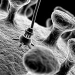
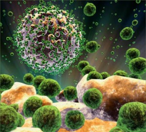
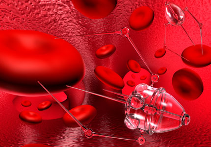
Drug delivery is the hotspot of research right now. Putting cancer therapy drugs into tumor areas is tricky. Nanobots are on the forefront of this area. Nanobots would theoretically detect cancer cells and destroy them effectively. Not only can nanobots be used for treating diseases, but they can be used for stopping aging, and interfering in a number of physiological functions for good use. They could also be used for cleaning pollution in the air, cleaning clothes, or anything else. The sky is the limit. Maybe nanobots could be used to create supercomputers and could be applied to electronic devices.
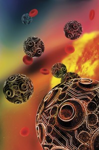
An artists perception of nanorobots.
Another method for deliviring drugs are vaults. Vaults are in almost all eukaryotic cells. They are made up of proteins, one MVP and two minor proteins, and some RNA. They appear to have no function, and are hollow inside. Vaults are shaped a little like hot dogs. However, they seem to have a lot of potential because they are not foreign in the body. Cancer drugs could be put inside the vaults and once in the cell, they can release the drug to destroy the cancer cell.
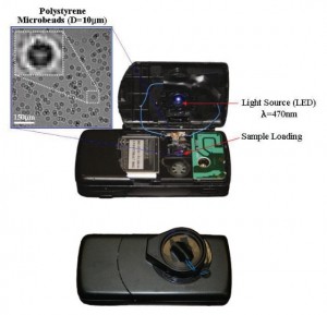
LUCAS is an easy way to view a blood sample by using a device that captures a shadow image of blood cells. It is cheap, easy, and fast. It could be used to diagnose HIV and malaria.
http://newsroom.ucla.edu/portal/ucla/cell-phone-prototype-of-lensless-77054.aspx
http://www.nano-biology.net/showabstract.php?pmid=16608733
http://www.ncbi.nlm.nih.gov/sites/entrez?db=pubmed&uid=14520404&cmd=showdetailview
http://www.physorg.com/news116071209.html
http://nano.cancer.gov/resource_center/sci_biblio_enabled-therapeutics_abstracts.asp
Posted in Uncategorized | Tagged Y |
Tuesday, July 7 2009
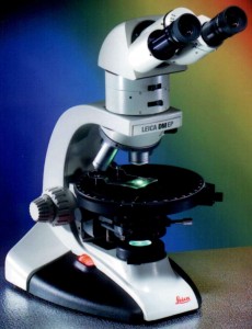
Microscopes come in all shapes and sizes, for different needs and purposes. Some cost millions, while others cost a few hundred bucks. They help see the tiny world around us, and in us. For example, there was a biomedical researcher using a scanning electron microscope (SEM) to look at tiny nanoparticle transporters. Pharmaceutical companies can use these nano devices for various purposes. They require microscopes to be observed to find stuff that could be wrong with it.
How do SEM microscopes work? First, an electrical current is passed through a metal, usually a tungsten filament. This is called an electron gun, and it fires electrons at the sample. Electromagnetic coils move the electrons with high speed and power. The electrons hit the sample and scatter back. Receptors take these electrons and send the information to a computer for analysis. Images can be magnified up to 2 million times in exceptional microscopes. SEMs are good for producing 3D images. However, samples must be viewed in a vacuum, therefore, any sample with water must undergo special preparation. They must be frozen at an extremely rapid rate so the water does not form crystals that can damage the sample. Therefore biological specimens that are alive are difficult to examine.
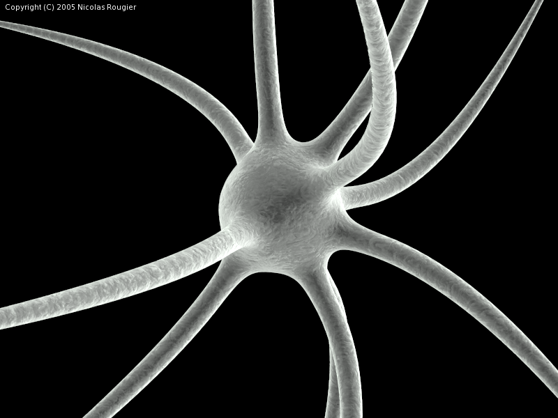
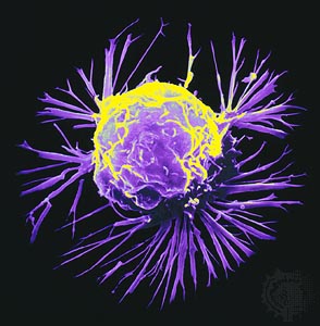
This is an image of a neuron under an SEM
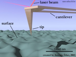
Another microscope used is an atomic force microscope (AFM). These microscopes have a cantelever, something that looks like a diving board and is very flexible. It is used for looking at the topography of the sample. They are handy for looking at live samples. They cantelever moves up and down along the surface. This movement is recorded by shooting a laser at the cantelever and then bouncing back to a photodiode. Resolution can be very high.
Which type of microscope is largely determined by need. Scientists will want to look for the cheapest method avaliable, because using some of the more advanced microscope techniques can be very costly.
http://www.mos.org/sln/SEM/
http://www.nanoscience.com/education/AFM.html
http://www.pubmedcentral.nih.gov/articlerender.fcgi?artid=537754
http://en.wikipedia.org/wiki/Scanning_electron_microscope
http://www.eetimes.com/news/semi/showArticle.jhtml?articleID=187002424
Posted in Uncategorized | Tagged Y |
Today, I asked myself what nanotechnology is. Is it the use of technology of microscopic proportions that will further society into the future? Or is it the study of minuscule organisms and the way humans will soon use them to further our race? It is all of those. Presently, nanotechnology is only in its start, but with every passing moment, the subject is evolving into something tangible and incredible. I never realized this, but nanotechnology has been in this world longer than most people alive today. Born in the mid 20th century, authors and scientists thought of infinitesimal robots that would assist the human race in everything. Ever since then, the subject was a growing subject that would soon be known across the world. This new science has affected many people especially scientists and artists. Nanotechnology allows a scientist to create new fields of study and creation. Nanotechnology has allowed scientists to think of new ways to cure diseases and explore the human body. For art, nanotechnology shows a new way of perceiving new things. Artists use nanotechnology to produce different forms of art, such as photographs of microscopic organisms and exhibits of new sciences to teach the public. Though many may say that science and art are totally different subjects and there is no way to connect the two subjects, the two are more alike than different. Science can be described as a new form of art and art can use different forms of science. They assist each other. All in all, nanotechnology has helped many think of new ways to cure disease and create easier ways to help humanity.
http://en.wikipedia.org/wiki/Nanotechnology
http://www.zyvex.com/nano/
http://science.howstuffworks.com/nanotechnology.htm
http://www.npr.org/templates/story/story.php?storyId=4111499
http://www.foresight.org/Updates/Update41/Update41.2.html
Posted in Uncategorized | Tagged group y |